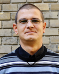
Nicolae LEOPOLD, Assoc. Prof., Ph.D. - Person in charge from Partner I
I was introduced into the field of Raman and surface-enhanced Raman spectroscopy (SERS) in the laboratory of Prof. W. Kiefer at the Institute of Physical Chemistry, University of Würzburg. After PhD, I had the opportunity to work for one year in the group of Prof. G. Gauglitz at the Institute of Physical Chemistry, University of Tübingen, in the field of optical chemical sensors and biosensors. My contribution to the development of microarray biochips, for sensory purposes can be found in the following book chapter: Mönch, W., et al., Microarray Biochips - Thousands of Reactions on a Small Chip (MOBA), in Biophotonics: Visions for Better Health Care. Wiley-VCH Verlag, 2006, p.405-476.
During an EIF Marie Curie fellowship at the Institute for Analytical Chemistry of the TU Vienna, in the group of Prof. B. Lendl, I developed analytical applications of SERS spectroscopy, showing the possibility of on-line SERS detection in a flow cell after capillary electrophoresis separation (N. Leopold and B. Lendl, Anal. Bioanal. Chem. 396, 2010, 2341 IF3.5). After my return, I developed at the BBU an approach for on-line SERS detection in a flow system after sequential injection of analytes (Herman et al. Anal. Bioanal. Chem. 400, 2011, 815 IF3.8). Concerning SERS detection of microorganisms, I collaborated with Dr. Dina and Prof. Haisch from TU München for single cell SERS detection of E. coli and P. mirabilis bacteria (Mircescu et al. Anal. Bioanal. Chem 406, 2014, 3051 IF3.6).
Since 2012, I perform research together with my students in the Raman-AFM laboratory at BBU http://www.phys.ubbcluj.ro/raman/index.htm, my present research being focused on the development of functional metallic nanoparticles as SERS substrate for protein investigation, and Raman mapping of haloarchaea, pathogen microorganisms and single human cells.
Research funding: 2006-2008 grant no. ET_91 (project director, 39.381 euro) | 2008-2010 grant no. RP_6 (project director, 135.769 euro) | 2010-2013 grant no. TE_323 (project director, 178.151 euro) | 2006-2008 Marie Curie Intra-European Fellowship grant no. MEIF-CT-2006-041451 (Fellow, 94.846 euro).
Metrics: Publications: 75; Citations: 1227 total citations; h-index: 14, Award of the Romanian Academy 2010, ORCID Profile; Google Scholar Profile
Webpage: http://www.phys.ubbcluj.ro/~nicolae.leopold
Email: nicolae.leopold@phys.ubbcluj.ro

Vasile CHIS, Full Prof., Ph.D. - Senior reseacher
I started my research carrier working on free radicals on gamma-irradiated amino acids aiming to get information on their geometrical and electronic structures. Working with prof. Marina Brustolon from Padova University I used cw and pulsed EPR as well as ENDOR spectroscopies as experimental techniques for measuring the hyperfine coupling tensors of the free radicals induced by gamma-irradiation of single crystals. Their structures have been then inferred indirectly by using quantum chemical methods. The main contribution to the field is reflected in the following publications: New Radical Detected by HF-EPR, ENDOR and Pulsed EPR in a Room Temperature Irradiated Single Crystal of Glycine; M. Brustolon, V. Chis, A.L. Maniero, L.C. Brunel; J. Phys. Chem. A, 101, 1997; Three different tyrosyl radicals identified in L-Tyrosine HCl crystals upon gamma-irradiation: magnetic characterization and temporal evolution, A. L. Maniero, V. Chis;, A. Zoleo, M. Brustolon, A. Mezzetti, J. Phys. Chem. B, 112, 2008.
After obtaining the PhD degree at BBU I became interested in studying the molecular and electronic structures of biomolecular systems like: amino acids, drugs and host-guest complexes for medical applications. Using experimental techniques (IR, Raman, NMR) coupled to computational chemistry approaches (DFT, MP2, semiempirical methods) we are able to fully understand the electronic structures of the investigated compounds and to describe subtle features related to their transformation between the solid and liquid state. Dispersion interactions and excited states of biomolecules are also investigated in my group: Distinguishing Binding Modes of a New Phosphonium Dye with DNA by Surface-Enhanced Raman Spectroscopy, S. Miljanic, A. Dijanosc, M. Novak, T.G. Deliqeorqiev, I. Crnolatac, V. Chis, and I. Piantanida, RSC Advances, 6, 2016; Surface mediated chiral interactions between cyclodextrins and propranolol enantiomers: a SERS and DFT study, R. Stiufiuc, C. Iacovita, G. Stiufiuc, E. Bodoki, V. Chis, C.M. Lucaciu, Phys. Chem. Chem. Phys., 17, 2015; Weakly bound PTCDI and PTCDA dimers studied by using MP2 and DFT methods with dispersion correction, M. Oltean, G.S. Mile, M. Vidrighin, N. Leopold, V. Chis, Phys. Chem. Chem. Phys., 15, 2013; Investigations of the supramolecular structure of individual diphenylalanine nano- and microtubes by polarized Raman microspectroscopy, B. Lekprasert, V. Korolkov, A. Falamas, V. Chis, C.J. Roberts, S.J.B. Tendler, I. Notingher, Biomacromolecules, 13, 2012.
Research funding: 2007-2008 Understanding Ionic Liquids as Novel Solvent in Green Chemistry; INTAS Ref. Nr. 06-1000031-10105, SEE-ERA.NET Pilot Joint Call (project manager) | 2007-2009 Molecular systems with applications in molecular electronics - Contract.nr.1485 (project manager) | 2010-2013 Bio-functional Nanoparticles for Development of new Methods of Imaging, Sensing, Diagnostic and Therapy in Biological Environment (NANOBIOFUN); Manager of Partner P5-from 2012.
Metrics: Publications: 90; Citations: 702 total citations; h-index: 14, Award of the Romanian Academy 2008.
Webpage: http://www.phys.ubbcluj.ro/~vasile.chis/
Email: vasile.chis@phys.ubbcluj.ro

László SZABÓ, Ph.D. - Postdoctoral researcher
Since 2012, I perform research together with my students and collaborators in the Raman-AFM laboratory at BBU http://www.phys.ubbcluj.ro/raman/index.htm my present research being focused on the development of functional metallic nanoparticles as SERS substrate for protein investigation, and Raman mapping of haloarchaea, pathogen microorganisms and single human cells.
Scientific research: optical spectroscopy (IR, Raman, SERS, UV-VIS), DFT calculations, studying chemical sensors and biosensors, determination of chemical residues in complex matrices and development of new ultra-sensitive Raman sensors, based on surface enhanced Raman spectroscopy (SERS), for the detection and quantification of heavy metal ions at ng/l concentrations; development of new conjugated nanoparticles for nano-biosenzing of ahterosclerosis and thrombosis by means of surface enhanced spatial offset Raman spectroscopy. I developed at the BBU an approach for on-line SERS detection in a flow system after sequential injection of analytes (Herman et al. Anal. Bioanal. Chem 400, 2011, 815).
Research funding: 2010-2012 grant nr. PD_445 (project director, 80.000 euro) | 2013-2016 grant nr. PN-II-RU-TE-2012-3-0227 (project director, 160.000 euro) | 2010-2013 grant nr. TE_323 (project manager, 178.151 euro) | 2007-2010 grant nr. PN II IDEI ID_501 (PhD student researcher, 250.000 euro) | 2006-2007 International Scholarship given by "World Federation of Scientists" c/o ICSC – WORLD LABORATORY, Lausanne, Switzerland (210 EURO/month).
Metrics: Over 30 articles in professional journals, including 25 in ISI journal, sum impact factor: 38,064, sum citations: 157 (1st June 2016), H-index: 8, i10-index: 7., Nr. of posters as co-author 46, international conferences (23)., Google Scholar Profile
Email: szabolaci81@gmail.com
Andrei STEFANCU - Student
I was introduced to Raman Spectroscopy by Professor N. Leopold in the Raman Laboratory, at the Babes-Bolyai University, where I am studying the distribution of carotenoids and chlorophylls in Hematococcus Pluvialis algae by Raman and fluorescence imaging. So, I became quite familiar with these optical imaging methods.
The main carotenoid we focused on during this study was astaxanthin. To detect this pigment we used the laser line 532 nm which is resonant with astaxanthin, this effect leading to a higher Raman signal intensity.
To detect chlorophylls we registered the fluorescent signal emitted by it, by using the 532 nm and 633 nm laser lines for excitation. The spectra obtained by Raman mapping were analyzed by Principal Component Analysis and we obtained the cell picture in false colors.
Another field of my research is related to Raman mapping of human cells. Recently, we contributed with an oral presentation to the National Conference of Biophysics, 2-4 June 2016, Cluj Napoca: N. Leopold, V. Moisoiu, A. Stefancu, Raman Imaging of Dental Follicle Mesenchymal Stem Cells, Book of Abstracts, p.57.
Email: andrei.stefancu16@yahoo.com


